Restore Vision with SurgiPartner Precision-Guided Vitrectomy Surgery
Experience Clear, Stable Vision with Advanced Retinal Microsurgery
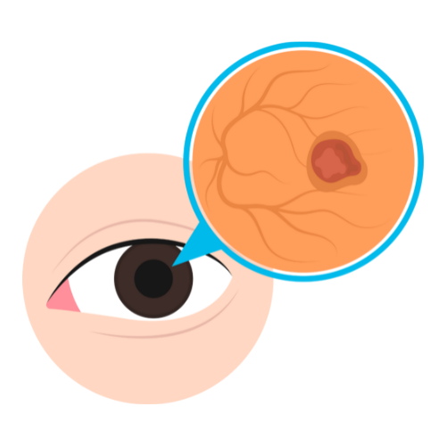
Vitrectomy Surgery in Hyderabad — Pars Plana Vitrectomy (PPV)
Vitrectomy — specifically Pars Plana Vitrectomy (PPV) — is a microsurgical procedure performed inside the eye to treat a range of sight-threatening retinal and vitreous conditions. SurgiPartner connects patients in Hyderabad with experienced vitreoretinal surgeons who specialise in both 23-gauge and 25-gauge small-gauge vitrectomy — advanced techniques offering faster recovery, less post-operative inflammation, and superior surgical precision.


What Is Vitrectomy Surgery?
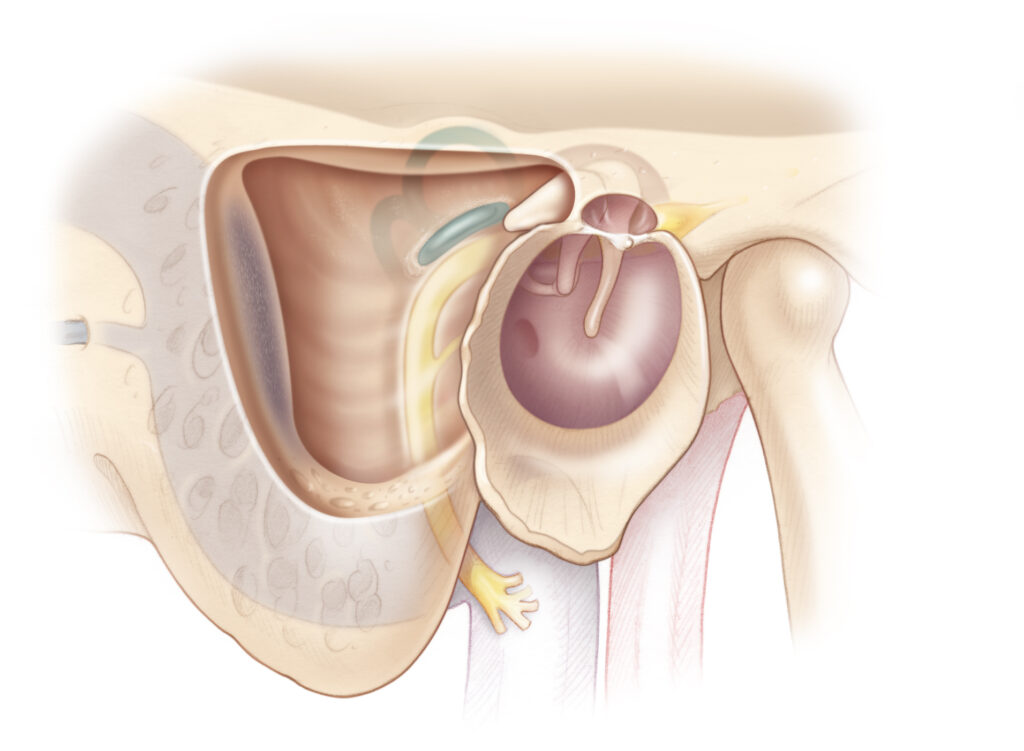
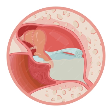
The vitreous is a clear, gel-like substance that fills the back two-thirds of the eye, maintaining its shape and allowing light to pass through to the retina. As we age, the vitreous liquefies and may pull away from the retina — a normal process called posterior vitreous detachment (PVD). In some cases, this separation creates tears in the retina, allows blood to enter the vitreous cavity, or leaves membranes on the retinal surface that distort vision.
Vitrectomy surgery removes the vitreous gel and any abnormal membranes, blood, or scar tissue from inside the eye, allowing the surgeon direct access to the retina to treat underlying conditions. The eye naturally replaces the removed vitreous with aqueous fluid over time, and in cases where tamponade (internal support) is needed, gas or silicone oil is temporarily placed inside the eye to hold the retina in position during healing.
Modern small-gauge vitrectomy (23g, 25g, 27g) uses micro-instruments through self-sealing incisions that do not require sutures, dramatically improving patient comfort and recovery compared to older techniques.
Conditions Treated With Vitrectomy Surgery
| Condition | How Vitrectomy Helps | Urgency |
|---|---|---|
| Retinal Detachment | Removes vitreous traction, flattens retina, allows laser or cryotherapy | Emergency — within 24–72 hours |
| Vitreous Haemorrhage | Removes blood-filled vitreous to restore clarity | Urgent / Elective |
| Diabetic Tractional RD | Removes membranes pulling retina away | Urgent |
| Macular Hole | Removes traction & ILM peeling for closure | Elective (3–6 months) |
| Epiretinal Membrane | Peels scar tissue improving vision | Elective |
| Endophthalmitis | Removes infected vitreous + antibiotics | Emergency |
| Dropped Nucleus | Retrieves lens material post surgery | Urgent |
| Intraocular Foreign Body | Removes foreign object from eye | Emergency |
What Is a Macular Hole? — One of the Most Common Vitrectomy Indications
A macular hole is a small, round opening in the macula — the central part of the retina responsible for sharp, detailed vision. It develops when the vitreous gel pulls on the macula as it separates from the retina during posterior vitreous detachment. Patients notice a central blind spot or distortion in their vision (metamorphopsia) — straight lines appear wavy or bent.
Macular holes are classified by size and stage on OCT imaging. Small macular holes (Stage 1–2) may occasionally close spontaneously; full-thickness macular holes (Stage 3–4) require surgical treatment for closure and vision recovery. Vitrectomy with ILM peeling and gas tamponade closes over 90% of macular holes in a single procedure.
What Is Epiretinal Membrane (Macular Pucker)?
An epiretinal membrane (ERM), also called macular pucker or cellophane maculopathy, is a thin layer of scar tissue that grows on the surface of the macula. As the membrane contracts, it wrinkles and distorts the macula, causing vision to appear blurred or wavy. Objects may appear distorted in size or shape. ERMs are common in adults over 50 and are associated with posterior vitreous detachment, prior retinal conditions, and intraocular surgery.
Treatment is surgical — vitrectomy with membrane peeling (using micro-forceps to carefully remove the ERM and often the underlying ILM) restores retinal anatomy in most cases. Vision improvement after ERM peeling occurs gradually over 3–12 months as the retina recovers its normal contour.
How Vitrectomy Surgery Is Performed — Step by Step
Step 1: Pre-Operative Preparation
Comprehensive retinal evaluation including OCT, fundus photography, B-scan ultrasonography (when media is cloudy), and fluorescein angiography as needed. Medical clearance for anaesthesia is obtained. Pupil-dilating drops are instilled to maximise intraoperative retinal visibility.
Step 2: Anaesthesia
Small-gauge vitrectomy can be performed under retrobulbar or peribulbar local anaesthesia with intravenous sedation (monitored anaesthesia care) or general anaesthesia. The choice depends on patient age, surgical complexity, and patient preference.
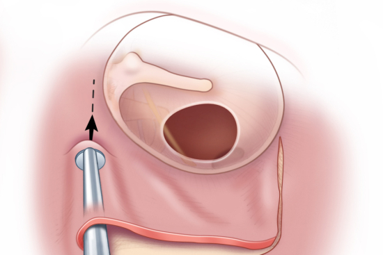
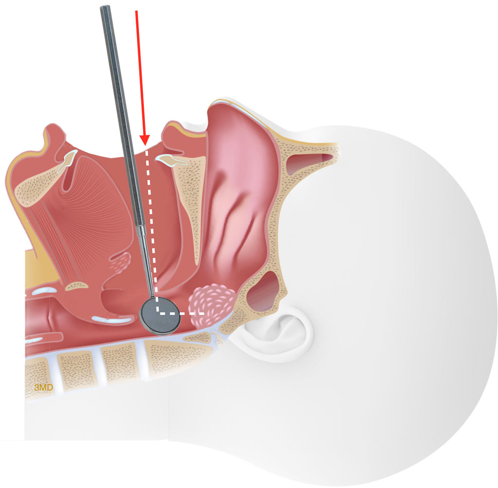
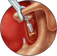
Step 3: Trocar Insertion (Small-Gauge Ports)
Three tiny self-sealing ports (25-gauge = 0.5mm diameter; 23-gauge = 0.6mm) are inserted into the pars plana — the flat part of the ciliary body — at 3.5–4mm from the limbus. Through these ports, the infusion cannula (maintaining eye pressure), the light pipe (illuminating the inside of the eye), and the vitreous cutter (removing the vitreous) are introduced.
Step 4: Core Vitrectomy
The vitreous cutter removes the vitreous gel at rates of 5,000–7,500 cuts per minute, minimising traction on the retina during removal. Surgical microscopy with wide-angle viewing systems gives the surgeon a panoramic view of the entire retinal surface.
Step 5: Condition-Specific Manoeuvres
Depending on the condition: retinal tears are treated with laser photocoagulation or cryotherapy; tractional membranes are removed with micro-forceps; blood from vitreous haemorrhage is aspirated; macular holes are treated with ILM peeling; foreign bodies are removed with intraocular forceps or magnets.
Step 6: Tamponade Placement
For retinal detachment and macular holes, the vitreous cavity is filled with a tamponade agent: short-acting gas (SF6, lasting 2–3 weeks), long-acting gas (C3F8, lasting 8–10 weeks), or silicone oil (lasting 3–6 months until removal). Tamponade holds the retina or macular hole in position during healing.
Step 7: Port Removal and Wound Closure
In small-gauge surgery, ports are self-sealing and typically do not require sutures. The conjunctiva is checked for integrity. The procedure typically takes 45 minutes to 3 hours depending on complexity.
Vitrectomy Recovery — What to Expect
a. Immediate: Eye patch applied; vision significantly blurred by gas bubble if used
- b. Head positioning: Critical for macular hole surgery and some retinal detachments — typically face-down for 1–2 weeks to position the gas bubble against the treated area
- c. No flying: Until all gas has absorbed (6–12 weeks for long-acting gas)
- d. Week 1: Eye drops commenced; review with surgeon; gas bubble visible and slowly decreasing
- e. Month 1: Gas absorbed; vision improving; retinal evaluation with OCT
- f. Month 1–6: Progressive visual recovery; glasses may need updating
- g. Silicone oil: If used, a second procedure to remove the oil is planned at 3–6 months
Frequently Asked Questions — Vitrectomy Surgery Hyderabad
Face-down positioning is required after vitrectomy with gas tamponade for macular holes — typically for 7–14 days post-operatively. The face-down position keeps the gas bubble directly against the macular hole, promoting closure. For retinal detachment with inferior or superior breaks, different positioning may be required depending on the location of the tamponade. Your surgeon will give precise positioning instructions b
ased on your specific procedure. Not all vitrectomies require face-down positioning — epiretinal membrane peeling, for example, does not.
Why Choose SurgiPartner for Vitrectomy Surgery?
SurgiPartner is a trusted destination for advanced vitrectomy surgery in Hyderabad, offering a combination of surgical excellence, advanced technology, and compassionate patient care.
01.
Expert Vitreoretinal Surgeons
Our surgeons have extensive experience in complex retinal microsurgery and advanced vitrectomy techniques.
02.
Advanced Retinal Technology
We utilize high-resolution OCT, digital retinal imaging, and state-of-the-art microsurgical platforms.
03.
Personalized Surgical Planning
Every vitrectomy procedure is tailored to the patient’s retinal condition and visual goals.
04.
Comprehensive Post-Operative Care
We provide structured recovery plans, continuous monitoring, and long-term retinal follow-up.
Book Your Consultation
Take the first step toward restoring and preserving your vision with SurgiPartner advanced vitrectomy surgery. Our retinal experts are committed to delivering precision-driven care with proven outcomes.
Advanced technology. Expert hands. Clearer vision.
What Our Patients Say

Posted onTrustindex verifies that the original source of the review is Google. I am thoroughly impressed with the SurgiPartner model, which delivers integrated, end-to-end patient support across the healthcare continuum—from initial consultations to treatment and surgical interventions, where necessary. I recently availed their Care Buddy service for a consultation, and the experience was highly seamless, efficient, and professionally managed. The structured Care Buddy support, combined with well-aligned financial assistance processes, significantly enhances the overall patient journey. This holistic and patient-centric approach effectively streamlines access to quality healthcare, making it more efficient and convenient in today’s dynamic environment.Posted onTrustindex verifies that the original source of the review is Google. I’m really impressed with the SurgiPartner concept. They provide end-to-end patient support—from doctor consultations to treatment and even surgery when required. I personally used their Care Buddy service for a consultation, and it was a smooth and positive experience. The Care Buddy assistance, along with support in financial processes, makes healthcare much more convenient, especially in today’s fast-paced lifestyle.Posted onTrustindex verifies that the original source of the review is Google. My Brother Arun Kumar and krishna from Bangalore. We are travelled to Hyderabad for LASIK surgery, and today we successfully underwent the procedure at American Laser Eye Hospital. The surgery went very smoothly and I am feeling very good after the procedure. The hospital environment is very clean and well maintained. All the staff and doctors are very polite, supportive, and professional. A special thanks to SurgiPartner CareBuddy for guiding and helping me throughout the entire process. From the beginning till the end of the day, they were with us at every step and provided excellent support. Overall, I had a very good experience and I highly recommend American Laser Eye Hospital for LASIK surgery with SurgiPartner support.Posted onTrustindex verifies that the original source of the review is Google. Really impressed with the concept of SurgiPartner. They offer complete patient support from doctor consultation to treatment and surgery if needed. I personally took a consultation through their Care buddy, and it was a great experience. The care buddy system and help with financial processes make healthcare much easier, especially in today’s busy world.Posted onTrustindex verifies that the original source of the review is Google. My name is Arun Kumar and I am from Bangalore. I traveled to Hyderabad for LASIK surgery, and today I successfully underwent the procedure at American Laser Eye Hospital. The surgery went very smoothly and I am feeling very good after the procedure. The hospital environment is very clean and well maintained. All the staff and doctors are very polite, supportive, and professional. A special thanks to SurgiPartner CareBuddy for guiding and helping me throughout the entire process. From the beginning till the end of the day, they were with us at every step and provided excellent support. Overall, I had a very good experience and I highly recommend American Laser Eye Hospital for LASIK surgery with SurgiPartner supportPosted onTrustindex verifies that the original source of the review is Google. Thank you for the support and service provided by SurgiPartner. The overall experience was smooth and the team was very helpful.Posted onTrustindex verifies that the original source of the review is Google. Staff is very good and nice receiving. They are very humble and their response is very good. Thank youPosted onTrustindex verifies that the original source of the review is Google. I want to thank Surgipartner for all the help and support during my LASIK surgery. From the beginning till the end, the team was very proactive with follow-ups and guided me clearly through every step, which made the whole experience easy and stress-free. I would especially like to appreciate CareBuddy Raju and Coordinator Anusha for their outstanding support. They were always in touch with both the hospital staff and me, helped reduce waiting time, and made sure everything went smoothly. Their care, quick responses, and constant guidance really made a big difference. Overall, I had a very positive experience and truly appreciate the support provided by Surgipartner.Verified by TrustindexTrustindex verified badge is the Universal Symbol of Trust. Only the greatest companies can get the verified badge who has a review score above 4.5, based on customer reviews over the past 12 months. Read more
Your Personalized Path to Wellness
Follow your step-by-step guide to a successful surgery and recovery, with our expert team supporting you all the way.
Book FREE Consultation
Fill in your details and we'll call you back to confirm your slot.