Say Goodbye to Breast Lumps with SurgiPartner Advanced Breast Care Solutions
Early Detection. Expert Care. Lasting Confidence.
Breast Lump Treatment in Hyderabad — Diagnosis, FNAC, Core Biopsy
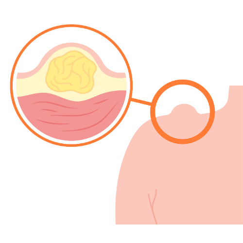
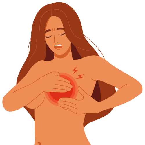
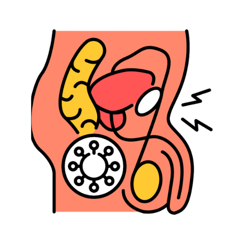
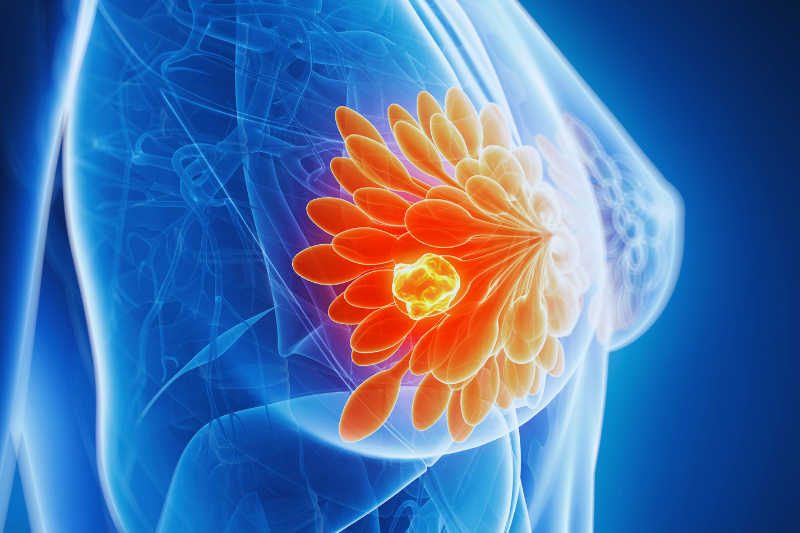
A breast lump is one of the most common reasons women seek medical consultation — and one of the most anxiety-provoking. The discovery of a breast lump, whether self-detected during routine self-examination or identified on screening imaging, requires prompt, thorough evaluation to determine whether it is benign or malignant. The reassuring fact is that the majority of breast lumps are benign — particularly in women under 40. SurgiPartner connects women in Hyderabad with breast surgeons and general surgeons for comprehensive, private breast lump assessment and treatment
The Triple Assessment — Essential Approach to Any Breast Lump
The international standard for evaluating any breast lump is the triple assessment — combining three complementary investigations that together provide near-complete diagnostic accuracy:
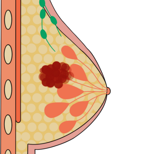
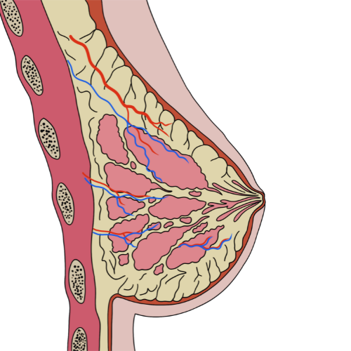
- Clinical assessment — careful history and physical examination by an experienced breast surgeon or general surgeon. Features assessed: age of the patient; duration and change in size of the lump; consistency (soft, firm, hard); shape (regular vs irregular); mobility (freely mobile vs tethered vs fixed to skin or chest wall); skin changes (dimpling, redness, peau d’orange — orange-peel skin texture); nipple changes (discharge, inversion, eczema — Paget’s disease); axillary lymph nodes (size, consistency, mobility). Each feature contributes to the clinical impression and urgency of further investigation.
- Imaging — the type depends on age and clinical findings: women under 35 undergo ultrasound (younger, denser breast tissue is poorly assessed by mammography); women 35 and over should have both ultrasound AND mammography. Ultrasound characterises the lump as cystic (fluid-filled) vs solid, and applies the BI-RADS classification to assess malignancy risk (BI-RADS 1–2: benign; BI-RADS 4–5: suspicious — biopsy required). MRI breast is used for high-risk screening, implant assessment, and pre-operative planning for known breast cancer.
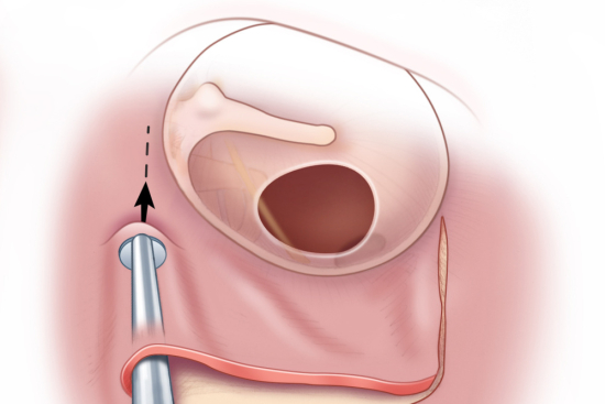
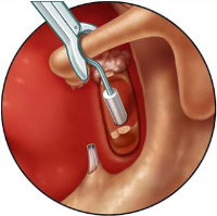
- Histology/Cytology — tissue sampling for microscopic diagnosis: FNAC (Fine Needle Aspiration Cytology) for simple cysts or clearly benign lesions; core needle biopsy (Tru-cut biopsy) for solid lumps requiring histological diagnosis (preferred — provides more tissue, architectural information, receptor status); vacuum-assisted biopsy for small or impalpable lesions.
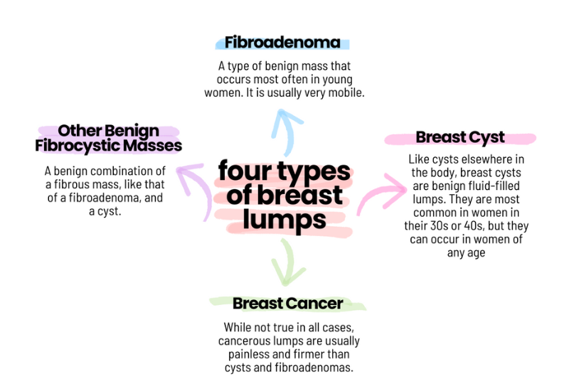
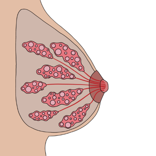
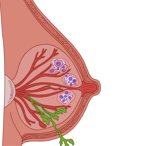
Types of Breast Lump — Differential Diagnosis
The international standard for evaluating any breast lump is the triple assessment — combining three complementary investigations that together provide near-complete diagnostic accuracy:
- Clinical assessment — careful history and physical examination by an experienced breast surgeon or general surgeon. Features assessed: age of the patient; duration and change in size of the lump; consistency (soft, firm, hard); shape (regular vs irregular); mobility (freely mobile vs tethered vs fixed to skin or chest wall); skin changes (dimpling, redness, peau d’orange — orange-peel skin texture); nipple changes (discharge, inversion, eczema — Paget’s disease); axillary lymph nodes (size, consistency, mobility). Each feature contributes to the clinical impression and urgency of further investigation.
- Imaging — the type depends on age and clinical findings: women under 35 undergo ultrasound (younger, denser breast tissue is poorly assessed by mammography); women 35 and over should have both ultrasound AND mammography. Ultrasound characterises the lump as cystic (fluid-filled) vs solid, and applies the BI-RADS classification to assess malignancy risk (BI-RADS 1–2: benign; BI-RADS 4–5: suspicious — biopsy required). MRI breast is used for high-risk screening, implant assessment, and pre-operative planning for known breast cancer.
- Histology/Cytology — tissue sampling for microscopic diagnosis: FNAC (Fine Needle Aspiration Cytology) for simple cysts or clearly benign lesions; core needle biopsy (Tru-cut biopsy) for solid lumps requiring histological diagnosis (preferred — provides more tissue, architectural information, receptor status); vacuum-assisted biopsy for small or impalpable lesions.
FNAC vs Core Needle Biopsy — Which Is Better?
FNAC (Fine Needle Aspiration Cytology) uses a fine needle (21–23 gauge) to aspirate cells from the lump — providing cytological (cell-level) diagnosis. Quick, minimally invasive, performed under local anaesthetic or no anaesthetic at all for simple cysts. Limitation: does not provide tissue architecture — cannot distinguish invasive from in-situ carcinoma; cannot provide hormone receptor status (essential for treatment planning); inadequate sample rate 10–15%.
Core needle biopsy uses a larger (14–16 gauge) spring-loaded needle to remove small core samples of tissue — providing histological (tissue-level) diagnosis. Performed under local anaesthetic with ultrasound guidance; takes 3–5 minutes. Provides definitive tissue diagnosis, architectural assessment, grade, receptor status (ER, PR, HER2 — essential for chemotherapy decision-making), and Ki67 proliferation index. The preferred technique for solid breast lumps at SurgiPartner partner hospitals.
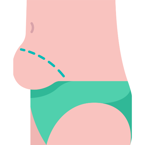
Surgical Treatment — Breast Lump Excision
All conducted with complete privacy and female coordinators available. Cashless insurance processing for all investigations and surgery. Call +91 9030053009.
Frequently Asked Questions — Breast Lump Treatment Hyderabad
Why Choose SurgiPartner?
At SurgiPartner, we combine advanced diagnostics, compassionate care, and precision surgery to ensure every woman receives the best treatment for breast lumps whether benign or cancerous.
01.
Expert Breast & Onco-Surgeons
Our specialists have vast experience in diagnosing and managing breast lumps, ensuring accurate detection and tailored treatment for every patient.
02.
Advanced Diagnostic & Surgical Technology
We use ultrasound-guided biopsies, mammography, and minimally invasive surgical methods to guarantee precision and safety.
03.
Personalized Treatment Plans
From simple cyst removal to complex lump excision, every procedure is customized to your age, health profile, and medical history - ensuring optimal outcomes.
04.
Compassionate, Confidential Care
Your comfort and privacy come first. Our female specialists provide a supportive, judgment-free environment throughout diagnosis and recovery.
Book Your Consultation
Take the first step toward peace of mind and a healthy future with SurgiPartner expert breast care team. Whether you’ve noticed a small lump or discomfort, early diagnosis leads to better outcomes.
Your health, your confidence, your choice – that’s the SurgiPartner promise.
What Our Patients Say

Posted onTrustindex verifies that the original source of the review is Google. I am thoroughly impressed with the SurgiPartner model, which delivers integrated, end-to-end patient support across the healthcare continuum—from initial consultations to treatment and surgical interventions, where necessary. I recently availed their Care Buddy service for a consultation, and the experience was highly seamless, efficient, and professionally managed. The structured Care Buddy support, combined with well-aligned financial assistance processes, significantly enhances the overall patient journey. This holistic and patient-centric approach effectively streamlines access to quality healthcare, making it more efficient and convenient in today’s dynamic environment.Posted onTrustindex verifies that the original source of the review is Google. I’m really impressed with the SurgiPartner concept. They provide end-to-end patient support—from doctor consultations to treatment and even surgery when required. I personally used their Care Buddy service for a consultation, and it was a smooth and positive experience. The Care Buddy assistance, along with support in financial processes, makes healthcare much more convenient, especially in today’s fast-paced lifestyle.Posted onTrustindex verifies that the original source of the review is Google. My Brother Arun Kumar and krishna from Bangalore. We are travelled to Hyderabad for LASIK surgery, and today we successfully underwent the procedure at American Laser Eye Hospital. The surgery went very smoothly and I am feeling very good after the procedure. The hospital environment is very clean and well maintained. All the staff and doctors are very polite, supportive, and professional. A special thanks to SurgiPartner CareBuddy for guiding and helping me throughout the entire process. From the beginning till the end of the day, they were with us at every step and provided excellent support. Overall, I had a very good experience and I highly recommend American Laser Eye Hospital for LASIK surgery with SurgiPartner support.Posted onTrustindex verifies that the original source of the review is Google. Really impressed with the concept of SurgiPartner. They offer complete patient support from doctor consultation to treatment and surgery if needed. I personally took a consultation through their Care buddy, and it was a great experience. The care buddy system and help with financial processes make healthcare much easier, especially in today’s busy world.Posted onTrustindex verifies that the original source of the review is Google. My name is Arun Kumar and I am from Bangalore. I traveled to Hyderabad for LASIK surgery, and today I successfully underwent the procedure at American Laser Eye Hospital. The surgery went very smoothly and I am feeling very good after the procedure. The hospital environment is very clean and well maintained. All the staff and doctors are very polite, supportive, and professional. A special thanks to SurgiPartner CareBuddy for guiding and helping me throughout the entire process. From the beginning till the end of the day, they were with us at every step and provided excellent support. Overall, I had a very good experience and I highly recommend American Laser Eye Hospital for LASIK surgery with SurgiPartner supportPosted onTrustindex verifies that the original source of the review is Google. Thank you for the support and service provided by SurgiPartner. The overall experience was smooth and the team was very helpful.Posted onTrustindex verifies that the original source of the review is Google. Staff is very good and nice receiving. They are very humble and their response is very good. Thank youPosted onTrustindex verifies that the original source of the review is Google. I want to thank Surgipartner for all the help and support during my LASIK surgery. From the beginning till the end, the team was very proactive with follow-ups and guided me clearly through every step, which made the whole experience easy and stress-free. I would especially like to appreciate CareBuddy Raju and Coordinator Anusha for their outstanding support. They were always in touch with both the hospital staff and me, helped reduce waiting time, and made sure everything went smoothly. Their care, quick responses, and constant guidance really made a big difference. Overall, I had a very positive experience and truly appreciate the support provided by Surgipartner.Verified by TrustindexTrustindex verified badge is the Universal Symbol of Trust. Only the greatest companies can get the verified badge who has a review score above 4.5, based on customer reviews over the past 12 months. Read more
Your Personalized Path to Wellness
Follow your step-by-step guide to a successful surgery and recovery, with our expert team supporting you all the way.
Book FREE Consultation
Fill in your details and we'll call you back to confirm your slot.