Diabetic Retinopathy Treatment in Hyderabad
Retinal detachment is a medical emergency. If you or someone you know is experiencing sudden flashes of light, a shower of floaters, or a shadow/curtain across your vision, seek immediate specialist care. SurgiPartner connects you with experienced retinal surgeons in Hyderabad within hours


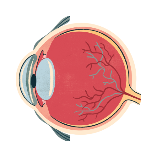
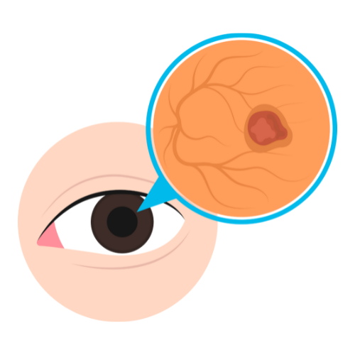
What Is Retinal Detachment?
The retina is the light-sensitive layer of tissue lining the back of the eye. It functions like the film in a camera — receiving visual images and transmitting them to the brain via the optic nerve. Retinal detachment occurs when this layer separates from its underlying supportive tissue, the retinal pigment epithelium (RPE), disrupting the blood supply and nutrient exchange the retina needs to function.
Without prompt treatment, retinal detachment leads to permanent, irreversible vision loss. The condition affects approximately 1 in every 10,000 people annually and is more common in individuals over 40, those with high myopia (nearsightedness), prior eye injuries, or a family history of retinal detachment.
The encouraging fact is that when diagnosed and treated early — ideally within 24–72 hours of symptom onset — retinal detachment surgery has a very high success rate in preserving and restoring vision.
Warning Signs of Retinal Detachment — Recognise the Emergency
Retinal detachment typically presents without pain, making the visual symptoms the only warning signals. Seek immediate medical attention if you experience:
- 1. Sudden onset of floaters — new, numerous dark spots, specks, or cobweb-like shapes in your vision that appear suddenly
- 2. Flashes of light (photopsia) — brief flashes of light in peripheral vision, particularly in dim light or darkness
- 3. Shadow or curtain — a dark shadow, veil, or curtain-like obstruction spreading across part of your visual field
- 4. Blurred central vision — if the detachment reaches the macula (central retina), central vision becomes blurred or distorted
- 5. Reduced peripheral vision — narrowing of the visual field from the sides
Important: Pre-retinal detachment warning signs include a sudden increase in floaters or new onset of flashes. These symptoms indicate a retinal tear — a precursor to detachment that can be treated much more simply before complete detachment occurs. Do not wait.
Types of Retinal Detachment
1. Rhegmatogenous Retinal Detachment (Most Common)
Caused by a tear or break in the retina that allows vitreous fluid to pass through and accumulate beneath the retina, separating it from the RPE. This is the most common type, accounting for approximately 90% of all retinal detachments. It is strongly associated with posterior vitreous detachment (PVD), high myopia, and prior eye trauma.
2. Tractional Retinal Detachment
Occurs when scar tissue or fibrovascular membranes on the retinal surface contract and pull the retina away from the RPE without a tear. This type is most commonly associated with proliferative diabetic retinopathy, sickle cell disease, and previous retinal surgery. It typically progresses more slowly than rhegmatogenous detachment.
3. Exudative (Serous) Retinal Detachment
Caused by fluid accumulating beneath the retina due to inflammation, tumours, or vascular disorders — without any tear or traction. Conditions such as posterior uveitis, choroidal tumours, and severe hypertension can cause this type. Treatment addresses the underlying cause rather than the retina directly.
Retinal Detachment Surgery — Treatment Options Explained
The choice of surgical technique depends on the type, location, and extent of detachment, as well as whether the macula is involved. SurgiPartner’s retinal specialists in Hyderabad use all established techniques and recommend the most appropriate approach for each patient.
1. Pneumatic Retinopexy
A gas bubble is injected into the vitreous cavity of the eye. The patient maintains a specific head position for several days, allowing the bubble to press against the retinal tear, closing it. Laser photocoagulation or cryotherapy seals the retina in place. Pneumatic retinopexy is suitable for small, superior retinal tears and can often be performed as an outpatient procedure. Success rates for appropriate cases are 70–80%, with repeat treatment available if needed.
2. Scleral Buckling
A flexible silicone band or sponge is placed around the circumference of the eye (sclera) and sutured in place. This indents the eye wall slightly, relieving the traction pulling the retina away and bringing the retina back into contact with the RPE. Cryotherapy or laser seals the retinal tear. Scleral buckling is particularly effective for younger patients with good lens clarity and superior or peripheral detachments. Success rates exceed 80–90% with experienced retinal surgeons.
3. Vitrectomy (Pars Plana Vitrectomy — PPV)
Vitrectomy is the most versatile surgical technique for retinal detachment. Three small incisions (0.5mm) are made in the eye, and micro-instruments remove the vitreous gel, release any tractional membranes, and allow the retina to be carefully flattened. The eye is then filled with a tamponade agent — either a gas bubble (SF6, C3F8) or silicone oil — that holds the retina in position while it reattaches. Laser photocoagulation seals the retinal tears. Vitrectomy is the preferred technique for complex or posterior detachments, macular involvement, and tractional retinal detachment.
What Happens If Retinal Detachment Is Left Untreated?
Untreated retinal detachment causes progressive, permanent vision loss. The sequence is predictable: a peripheral detachment that spares the macula initially preserves central vision, but as the detachment extends to reach the macula — typically within days to weeks — irreversible central vision loss occurs. Total retinal detachment leads to complete blindness in the affected eye, which cannot be reversed even with subsequent surgery. This is why retinal detachment is treated as an ophthalmic emergency requiring surgery within 24–72 hours of diagnosis.
Retinal Detachment Surgery — Recovery and Expectations
| Post-Surgery Phase | What to Expect | Important Instructions |
|---|---|---|
| First 24 hours | Eye patched; significant visual blurring; some discomfort | Strict head positioning if gas bubble used |
| Days 2–7 | Patch removed; vision slowly improving; gas bubble visible | No flying (gas expands at altitude — risk of blindness) |
| Week 2–4 | Gas bubble gradually absorbed; vision improving | No strenuous activity; avoid eye rubbing |
| Month 1–3 | Significant visual recovery; final result emerges | Regular follow-up with retinal surgeon |
| Month 3–12 | Vision continues stabilising; new glasses may be needed | Report any new floaters or vision changes immediately |
Important: If silicone oil was used as tamponade, a second surgery to remove the oil is typically planned 3–6 months after the initial procedure.
Factors That Affect Visual Outcome After Retinal Detachment Surgery
- 1. Macula involvement — the single most important factor. When the macula (central retina) is detached, even successful surgery may not restore 100% central vision. Macular-on detachments (macula still attached) have significantly better visual prognoses
- 2. Duration of detachment — shorter duration = better outcomes. Surgery within 24 hours of macular involvement is critical
- 3. Extent of detachment — localised detachments have better outcomes than total detachments
- 4. Surgeon experience — retinal surgery requires high-level subspecialty expertise; SurgiPartner only partners with experienced vitreoretinal surgeons
- 5. Patient age and overall eye health — younger patients with otherwise healthy eyes generally recover better visual acuity
Frequently Asked Questions — Retinal Detachment Surgery Hyderabad
Yes — retinal detachment is a medical emergency that requires surgery as soon as possible, ideally within 24–72 hours of symptom onset. When the macula (central retina) becomes detached, every hour of delay reduces the chance of recovering good central vision. If you have sudden new floaters, flashes of light, or a shadow in your vision, contact SurgiPartner immediately on +91 9030053009.
Visual recovery depends primarily on whether the macula was involved and how long the detachment was present before surgery. If the macula was not detached (macula-on detachment), most patients recover near-normal vision after successful surgery. If the macula was detached, central vision recovery is variable and may be incomplete even after anatomically successful surgery. Peripheral vision recovery is generally very good in both cases.
Why Choose SurgiPartner?
Choosing SurgiPartner means trusting your vision with experts in advanced retinal detachment surgery in Hyderabad.
01.
Expert Retinal Surgeons
Our specialists have extensive experience in complex retinal surgeries with high success rates.
02.
Advanced Surgical Technology
We use FDA-approved retinal imaging and microsurgical systems for unmatched precision.
03.
Personalized Treatment Plans
Each patient receives a customized surgical and recovery plan based on retinal health.
04.
Safe & Compassionate Care
From emergency diagnosis to post-surgery follow-ups, we ensure complete eye care support.
Book Your Consultation
Protect your eyesight with SurgiPartner’s advanced retinal detachment surgery in Hyderabad. Early intervention can save vision and restore clarity.
Clear vision isn’t a hope—it’s a SurgiPartner commitment.
What Our Patients Say

Posted onTrustindex verifies that the original source of the review is Google. I am thoroughly impressed with the SurgiPartner model, which delivers integrated, end-to-end patient support across the healthcare continuum—from initial consultations to treatment and surgical interventions, where necessary. I recently availed their Care Buddy service for a consultation, and the experience was highly seamless, efficient, and professionally managed. The structured Care Buddy support, combined with well-aligned financial assistance processes, significantly enhances the overall patient journey. This holistic and patient-centric approach effectively streamlines access to quality healthcare, making it more efficient and convenient in today’s dynamic environment.Posted onTrustindex verifies that the original source of the review is Google. I’m really impressed with the SurgiPartner concept. They provide end-to-end patient support—from doctor consultations to treatment and even surgery when required. I personally used their Care Buddy service for a consultation, and it was a smooth and positive experience. The Care Buddy assistance, along with support in financial processes, makes healthcare much more convenient, especially in today’s fast-paced lifestyle.Posted onTrustindex verifies that the original source of the review is Google. My Brother Arun Kumar and krishna from Bangalore. We are travelled to Hyderabad for LASIK surgery, and today we successfully underwent the procedure at American Laser Eye Hospital. The surgery went very smoothly and I am feeling very good after the procedure. The hospital environment is very clean and well maintained. All the staff and doctors are very polite, supportive, and professional. A special thanks to SurgiPartner CareBuddy for guiding and helping me throughout the entire process. From the beginning till the end of the day, they were with us at every step and provided excellent support. Overall, I had a very good experience and I highly recommend American Laser Eye Hospital for LASIK surgery with SurgiPartner support.Posted onTrustindex verifies that the original source of the review is Google. Really impressed with the concept of SurgiPartner. They offer complete patient support from doctor consultation to treatment and surgery if needed. I personally took a consultation through their Care buddy, and it was a great experience. The care buddy system and help with financial processes make healthcare much easier, especially in today’s busy world.Posted onTrustindex verifies that the original source of the review is Google. My name is Arun Kumar and I am from Bangalore. I traveled to Hyderabad for LASIK surgery, and today I successfully underwent the procedure at American Laser Eye Hospital. The surgery went very smoothly and I am feeling very good after the procedure. The hospital environment is very clean and well maintained. All the staff and doctors are very polite, supportive, and professional. A special thanks to SurgiPartner CareBuddy for guiding and helping me throughout the entire process. From the beginning till the end of the day, they were with us at every step and provided excellent support. Overall, I had a very good experience and I highly recommend American Laser Eye Hospital for LASIK surgery with SurgiPartner supportPosted onTrustindex verifies that the original source of the review is Google. Thank you for the support and service provided by SurgiPartner. The overall experience was smooth and the team was very helpful.Posted onTrustindex verifies that the original source of the review is Google. Staff is very good and nice receiving. They are very humble and their response is very good. Thank youPosted onTrustindex verifies that the original source of the review is Google. I want to thank Surgipartner for all the help and support during my LASIK surgery. From the beginning till the end, the team was very proactive with follow-ups and guided me clearly through every step, which made the whole experience easy and stress-free. I would especially like to appreciate CareBuddy Raju and Coordinator Anusha for their outstanding support. They were always in touch with both the hospital staff and me, helped reduce waiting time, and made sure everything went smoothly. Their care, quick responses, and constant guidance really made a big difference. Overall, I had a very positive experience and truly appreciate the support provided by Surgipartner.Verified by TrustindexTrustindex verified badge is the Universal Symbol of Trust. Only the greatest companies can get the verified badge who has a review score above 4.5, based on customer reviews over the past 12 months. Read more
Your Personalized Path to Wellness
Follow your step-by-step guide to a successful surgery and recovery, with our expert team supporting you all the way.
Book FREE Consultation
Fill in your details and we'll call you back to confirm your slot.