Scar-Minimizing Techniques • Quick Recovery • Expert Dermatologists & Surgeons
Safe & Advanced Mole Removal Treatment
Mole Removal in Hyderabad — Safe Surgical Excision, Laser Treatment
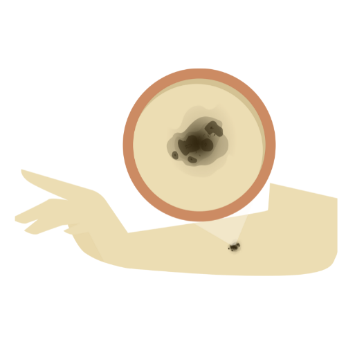
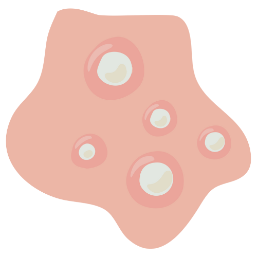
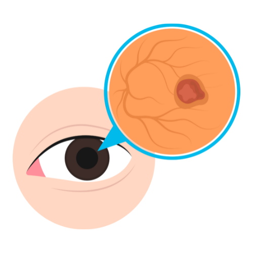
Moles (melanocytic naevi) are common benign pigmented lesions caused by clusters of melanocytes (pigment-producing cells) in the skin. While the vast majority are completely harmless, certain moles — particularly those that are changing, irregular, or symptomatic — may require removal either for medical (melanoma screening) or cosmetic reasons. SurgiPartner connects patients in Hyderabad with dermatologists and plastic surgeons for expert mole assessment and safe removal.


When Is Mole Removal Necessary? — The ABCDE Rule
Any mole displaying the following warning features (ABCDE criteria) requires urgent dermatological assessment and consideration of surgical excision with histopathology
| Type | Composition | Key Clinical Features | Treatment Consideration |
|---|---|---|---|
| Simple lipoma | Mature fat cells with thin capsule | Most common; soft; non-tender; slow growth; single lesion | Standard excision or minimal incision technique |
| Angiolipoma | Fat cells + prominent vascular component | Often painful or tender; may be multiple; typically <3cm; forearm most common site | Excision provides permanent pain relief; all specimens sent for histopathology |
| Fibrolipoma | Fat + fibrous stroma in varying proportions | Firmer consistency than simple lipoma; less compressible; can be confused with other lesions clinically | Requires standard excision; harder to enucleate than simple lipoma |
| Myelolipoma | Fat + haematopoietic (bone marrow-like) tissue | Most commonly adrenal; incidentally found on imaging; rarely subcutaneous | Adrenal myelolipoma managed by endocrinology/urology; subcutaneous excised with histopathology |
| Pleomorphic lipoma | Fat + giant cells with floret nuclei | Neck/shoulder area; elderly men; clinically indistinguishable from simple lipoma | Excision essential; histopathology confirms benign nature and excludes atypical lipomatous tumour |
| Spindle cell lipoma | Fat + spindle cells + ropey collagen | Neck/shoulder/back; men 50–70 years; clinically benign but requires histological diagnosis | Excision + histopathology; MDM2 mutation analysis in selected cases |
Additional warning signs: bleeding from a mole without trauma, itching, crusting, ulceration, or a new dark streak in a fingernail.


Mole Removal Techniques
Surgical Excision with Histopathology (Recommended for Suspicious Moles)
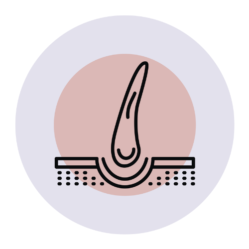
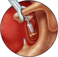
The gold standard for any mole with suspicious features. An elliptical excision removes the mole with a defined margin of normal surrounding skin. The specimen is sent to histopathology — the only definitive way to exclude melanoma. Performed under local anaesthesia. Leaves a fine linear scar; heals within 2–3 weeks. This is the only technique that provides a definitive diagnosis.
Shave Biopsy / Shave Excision
For raised (pedunculated or dome-shaped) benign-appearing moles. A scalpel or razor shaves the mole flush with the skin surface. Specimen submitted for histopathology. Not recommended for flat moles or suspicious lesions due to inadequate depth assessment. Leaves a flat, round scar that usually heals well.
Laser Mole Removal
CO₂ or Q-switched laser ablates superficial mole tissue. Advantages include no incision and minimal scarring for small flat moles. Critical limitation: no specimen for histopathology — laser mole removal should only be used for clearly benign, dermatoscopically assessed moles. Never appropriate for suspicious or changing moles. Flat, flesh-coloured or lightly pigmented small moles in cosmetically sensitive areas (face) are the best candidates.
💡 Important: SurgiPartner’s partner dermatologists perform dermoscopy (skin surface microscopy) of all moles before removal to assess benignancy. Any suspicious mole is always removed surgically with histopathology — never by laser alone. Call +91 9030053009 .
Frequently Asked Questions — Mole Removal Hyderabad
All mole removal techniques leave some degree of mark or scar, though the appearance varies significantly by technique and location. Surgical excision leaves a fine linear scar that is typically 2–3× the diameter of the mole; this fades to a faint pale line within 6–12 months with good scar care. Shave excision leaves a flat, round, slightly depigmented mark that blends well with surrounding skin. Laser treatment leaves minimal to no visible mark for small, flat moles. Cosmetically sensitive areas (face, neck) are approached with meticulous technique and fine sutures by SurgiPartner's plastic surgeons. Silicone scar management from week 3 optimises all scar outcomes.
Why Choose SurgiPartner for Mole Removal?
Expert dermatologists using advanced, scar-minimizing techniques for safe, precise, and cosmetically superior results.
01.
Experienced Dermatologists & Surgeons
Specialists trained in cosmetic and medical skin procedures.
02.
Advanced Scar-Minimizing Techniques
Modern technology ensures precision and better cosmetic results.
03.
Safe & Hygienic Environment
Strict sterilization and safety protocols followed.
04.
Complete Aftercare Support
Follow-ups, biopsy reports (if needed), and skin care guidance.
Book Your Consultation
Take the first step toward relief from abdominal pain with SurgiPartner Advanced Appendectomy Surgery.
Live free from pain and discomfort that’s the SurgiPartner promise.
Patient Experiences – Mole Removal
“The mole on my face is gone without any visible scar.”
Your Personalized Path to Wellness
Follow your step-by-step guide to a successful surgery and recovery, with our expert team supporting you all the way.
Book FREE Consultation
Fill in your details and we'll call you back to confirm your slot.